Example FRCS Plast questions
25 free FRCS(Plast) example questions
Free single best answer (SBAs) questions spanning the FRCS(Plast) curriculum, from hand and lower limb trauma to head and neck, breast, burns, skin and aesthetics. Every question is written, illustrated and referenced to the standard of the real exam, with no sign-up required.

The pathology is isolated to the skin envelope: dermatochalasis without steatoblepharon, lid laxity, tear trough deformity, or midface descent. The component-based approach to lower blepharoplasty dictates intervention only on the abnormal layer - here, a subciliary skin-pinch excision that preserves the orbicularis oculi, the orbital septum, and the lower-lid retractors.
This minimises the principal causes of postoperative lid malposition:
Orbicularis denervation
Middle-lamellar scarring, and
Septal violation.
Other answers:
Option A removes fat she does not have and leaves the skin untouched.
Option B is overtreatment that introduces denervation risk and is unnecessary in the absence of fat or muscle pathology.
Option D adds canthal support in a lid with a normal snap-back and good tarsoligamentous tone - overtreatment again.
Option E is reserved for significant midface descent with tear trough deformity, neither of which is present.

Two questions decide the operation:
How lax is the lid?
Distraction of 8 mm (normal <6 mm) and a lid that will not snap back without a blink mean marked laxity - a simple support stitch will not be enough.
Where is the laxity?
Pulling the lid laterally tightens the canthal tendon. If the laxity were in the tendon, this would take up the slack. Here, redundancy persists and the canthal angles stay sharp; the excess is in the lid itself, so tissue must be excised: full-thickness horizontal shortening (modified Kuhnt-Szymanowski), with lateral canthal support.
Other answers:
A - Lateral tarsal strip alone: tightens a lax canthal tendon, the commoner problem in the ageing lid, but this lid stayed redundant even when the tendon was tightened, so a strip alone leaves excess behind.
B - Canthopexy: a plication stitch without tendon division; supports mild-to-moderate laxity or prevents malposition during routine blepharoplasty. It excises no excess tissue.
D - Mid-lid wedge without canthal support: removes tissue but leaves the lateral canthus unsupported - with risk of recurrence and canthal rounding.
E - Orbicularis sling: a suspension for the paralytic lid in facial palsy; it does not shorten a lax lid.

Acanthosis means epidermal hyperplasia - thickening of the stratum spinosum, usually with elongated rete ridges. It is a non-specific reactive change, so the diagnosis rests on the pattern of atypia, not the acanthosis itself.
Full-thickness keratinocyte atypia with an intact basement membrane and no dermal invasion defines squamous cell carcinoma in situ (Bowen disease). Extension down follicular infundibula is characteristic and explains why superficial destructive treatments sometimes fail.
Other answers:
Hypertrophic actinic keratosis - atypia confined to the lower third, alternating ortho- and parakeratosis sparing the adnexal ostia.
Verruca vulgaris - papillomatosis (increased depth of corrugations at the dermoepidermal junction), koilocytes, tiers of parakeratosis over papillary tips.
Superficial BCC - buds of basaloid cells with peripheral palisading (nuclei oriented perpendicular to the stroma) and retraction clefting.
Extramammary Paget disease - pagetoid spread: large pale cells scattered singly through the epidermis, CK7 and PAS positive.
Lower-leg Bowen disease heals poorly. BAD guidance supports 5-fluorouracil, cryotherapy, curettage, imiquimod or photodynamic therapy, reserving excision for diagnostic uncertainty, treatment failure or suspected invasion.
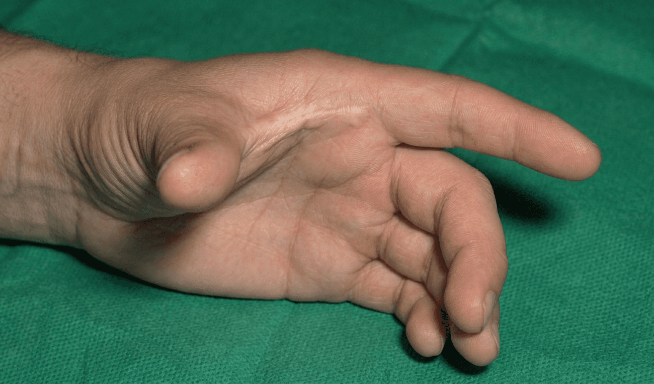
The five-flap plasty, "jumping-man" flap, combines two opposing Z-plasties with a central V-Y advancement.
It is the technique of choice here:
The paired Z-plasties break up and lengthen the band using supple lateral skin, whilst the central V-Y advances the lax proximal palmar skin directly into the apex of the web. F
Flaps marked A–E transpose to B-A-C-E-D.
Other answers:
Four-flap plasty - gives the greatest theoretical deepening for a given central limb (Fraulin & Thomson) but requires greater transverse laxity for flap transposition and has no central advancement, less suited when dorsal laxity is limited.
Release and grafting is reserved for severe, broad contractures where local tissue is insufficient.
A single Z-plasty - lengthens (~75% for 60° limbs) but cannot adequately deepen a web.
V-Y advancement alone - does not address the band.


The most likely diagnosis is Stewart–Treves syndrome (STS) - cutaneous angiosarcoma arising in a chronically lymphoedematous limb. Long-standing post-mastectomy arm lymphoedema with ~14-year latency, multifocal violaceous lesions confined to the affected arm, and progressive coalescence over months is classical.
Each distractor has a discriminating feature:
Radiation-induced angiosarcoma (B) is the closest histological mimic - both are MYC-amplified angiosarcomas, but anatomically arises within the irradiated field (chest wall or residual breast skin), not in the lymphoedematous arm. Mean latency is also shorter (~5–10 years).
Kaposi sarcoma (A) produces violaceous lesions but is HHV-8-driven, occurring in immunosuppressed/HIV-positive patients - not chronic post-mastectomy lymphoedema.
Atypical vascular lesion (C) is a benign post-radiation reactive lesion: typically small, static, MYC-negative, and lacks the progressive, multifocal, coalescent behaviour described.
Cutaneous breast metastases (E) typically present on the chest wall, not as multifocal violaceous lesions in the lymphoedematous arm.
Diagnosis is confirmed by incisional biopsy: CD31, CD34 and ERG positivity with MYC amplification on FISH.
Management is via the sarcoma MDT - wide local excision with clear margins, with amputation reserved for extensive proximal disease. Prognosis is poor: median survival ~19–31 months, 5-year survival ~10–15%.

There are no clinical signs of infection - no cellulitis, no purulence, systemically well; therefore MRSA on the swab represents colonisation of an open wound, not a surgical site infection.
NICE is clear that microbiology results alone should not drive antibiotic prescribing; treat the patient, not the swab. Systemic anti-MRSA agents (doxycycline, teicoplanin) are reserved for clinical infection.
T-junction breakdown is the commonest wound complication of Wise-pattern reduction, arising from tension and relative ischaemia where three flaps meet. These are usually minor problems that heal by secondary intention with simple dressings and rarely need surgical revision; early re-excision and closure recreates the same tension and typically breaks down again.
Decolonisation remains worthwhile. Standard UK regimens are nasal mupirocin 2% three times daily for 5 days plus a daily chlorhexidine 4% (or octenidine) body wash, arranged with infection prevention and the GP. The CLEAR trial showed post-discharge decolonisation reduced subsequent MRSA infection by around 30%.
Mupirocin should not be applied to the wound bed - that drives mupirocin resistance and is not a decolonisation regimen. Document MRSA status for any future implant-based or revision surgery, and advise on occupational health review before returning to work.
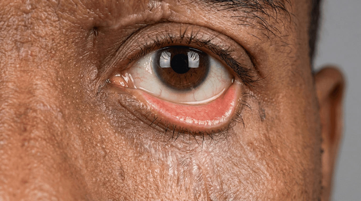
Ectropion is classified by what has failed:
Involutional - horizontal laxity of the tarsoligamentous sling; ageing. Corrected by shortening (lateral tarsal strip).
Cicatricial - vertically short anterior lamella; burns, trauma, actinic damage, over-resection at blepharoplasty. Corrected by releasing and replacing skin.
Paralytic - loss of orbicularis tone; facial nerve palsy. Corrected by static support (tarsal strip, sling, tarsorrhaphy).
Mechanical - the lid is dragged down by weight; tumour, oedema. Corrected by removing the cause.
This patient has both cicatricial and involutional elements - the lid is tethered and cannot be lifted above the pupil, and it distracts 9 mm with a delayed snap-back. Correcting only one will recur.
Release the scar through a subciliary incision until the lid lifts freely, resurface with an oversized full-thickness graft (upper lid, pre- or post-auricular, supraclavicular), then tighten with a lateral tarsal strip. Support the lid with a Frost suture and bolster.
Other answers:
A - tightens but leaves the scar.
B - Split-thickness grafts contract, which is what caused the initial problem.
C - adjusts the midface without adding skin.
E - suits an immature, untethered scar, not established keratopathy.

Laser Doppler imaging (LDI) measures dermal microvascular blood flow, which correlates with burn depth and, more usefully, healing potential.
NICE recommends LDI as an adjunct to clinical assessment in burns of indeterminate depth. Accuracy is highest between days 2 and 5 post-burn, which is why very early scanning is unreliable.
Low flux (<220 PU, blue) predicts healing beyond 21 days, indicating deep dermal or full-thickness injury. Wounds taking longer than 21 days to heal carry a high risk of hypertrophic scarring, so the standard of care is early tangential excision and grafting, particularly in functionally important areas such as the dorsum of the hand.
Other answers:
Continuing dressings or repeating the scan delays definitive treatment.
Biobrane is suited to confirmed superficial partial-thickness burns.
Biopsy is invasive, samples only one point, and adds nothing once LDI has predicted non-healing.
The phenotype - tall stature, arachnodactyly proxy (arm span > height), high-arched palate, progressive pectus excavatum, and a maternal history of aortic surgery, strongly suggests Marfan syndrome.
Pectus excavatum occurs in approximately two-thirds of Marfan patients and may be the presenting feature; missing the underlying connective tissue disorder risks fatal aortic dissection. Before any chest wall intervention, the candidate must undergo:
Echocardiography (aortic root, mitral valve prolapse)
Slit-lamp examination for ectopia lentis, and
Revised Ghent criteria assessment via clinical genetics
Other options:
Option A is wrong because surgery without ruling out aortopathy is unsafe and recurrence rates after pectus repair are higher in Marfan.
Option B misrepresents current NHS England policy: since the 2023 interim commissioning statement, pectus surgery is commissioned where the Haller index exceeds 3.25 and there is documented cardiorespiratory compromise (cardiac compression on MRI, arrhythmia, or dysphagia).
Options D and E address the deformity in isolation and miss the systemic diagnosis.

The third part of the axillary artery gives off the subscapular artery, its largest branch. This divides almost immediately into two:
Circumflex scapular artery
Thoracodorsal artery
The circumflex scapular turns backwards through the triangular space to reach the scapula. In contrast, the thoracodorsal keeps heading downwards, running with the thoracodorsal nerve, and enters the deep surface of latissimus dorsi about 8 to 14 cm beyond the split. The arrow follows that downward course, so it marks the thoracodorsal.
The other two branches sit elsewhere:
Lateral thoracic - descends over the chest wall, much further medially.
Posterior circumflex humeral - heads laterally through the quadrangular space with the axillary nerve.
NB - Option E is incorrect for two reasons: the subscapular comes off the third part, not the second, and the arrow lies beyond the point where it divides.
Lose this artery and you lose the dominant pedicle of the latissimus dorsi flap - although the flap may still survive on retrograde flow through the serratus branch!
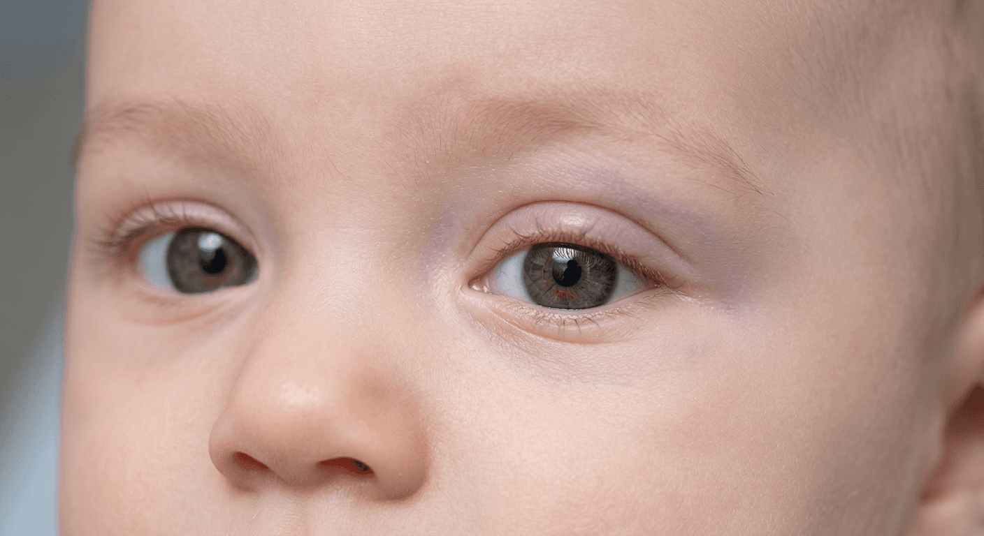
The periocular region is a high-risk site because the amblyogenic window is the first year of life.
Three concerns are present:
Deprivation from ptosis
Strabismus
Astigmatism - from mass effect on the globe.
A clear visual axis is falsely reassuring. A deep lesion indenting the globe is amblyogenic in its own right, so watchful waiting is wrong despite the natural history of involution; even one to two weeks of visual deprivation under the age of two can produce permanent cortical visual deficit.
Oral propranolol is first-line systemic therapy. The licensed regimen of 3 mg propranolol base/kg/day for six months achieved complete or near-complete resolution in 60.4% versus 3.6% for placebo. Efficacy is greatest during the proliferative phase, so referral should be urgent. Check baseline heart rate and blood pressure, and screen for PHACE if the lesion is segmental and over 5 cm. Joint paediatric ophthalmology input for refraction, spectacles and occlusion is mandatory - treating the haemangioma does not treat established amblyopia.
Other answers:
Topical timolol suits thin superficial lesions and will not penetrate a deep one.
Pulsed dye laser addresses colour, not bulk.
Intralesional triamcinolone risks embolic retinal artery occlusion and eyelid necrosis, and is largely superseded.
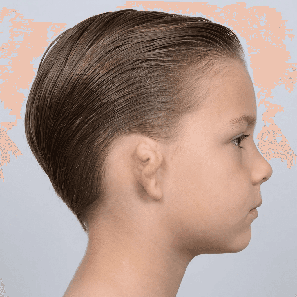
Timing is driven by costal cartilage volume, not chronological age or auricular growth.
The auricle reaches roughly 85% of adult size by 4 years and near-adult vertical height by 6 - which is why Brent operates from about 6, his multi-stage framework requiring less cartilage. Nagata's two-stage technique needs a bulkier three-dimensional framework, with thresholds of age 10 and chest circumference ≥60 cm at the xiphoid. This boy is 7 but measures 51 cm, so an adult-sized framework cannot be reliably fabricated. An undersized construct gives a small, poorly defined ear that is difficult to salvage. Monitor growth, time surgery to cartilage availability, and support him psychologically at school meanwhile.
Reconstruction demands virgin, unscarred skin and an intact superficial temporal artery, so autologous reconstruction precedes any atresiaplasty. In unilateral microtia with normal contralateral hearing, UK practice generally favours a bone-conduction device over canal surgery. Switching framework material purely to work around a small chest abandons the family's counselled choice.
Ordinal data consist of ordered categories where the rank is meaningful, but the intervals between successive categories are not necessarily equal.
House–Brackmann grade (I–VI) is the classic surgical example - Grade III is worse than Grade II, but the clinical "distance" from Grade II to III is not the same as from Grade V to VI.
Classifying the others:
Age and operative time are continuous (ratio) - measured on a numerical scale with equal intervals and a true zero.
Sex is nominal (dichotomous / binary) - unordered categories.
Aetiology is nominal (categorical, polytomous) - multiple unordered categories.
The practical implication:
Ordinal data should not be analysed with parametric tests that assume equal intervals (e.g. t-test, Pearson's r).
Appropriate methods include - Mann–Whitney U test (two groups), Kruskal–Wallis (>2 groups), Wilcoxon signed-rank (paired), and Spearman's rank correlation.
Other ordinal scales worth recognising include: Sunderland nerve injury grading, Gustilo–Anderson, Clavien–Dindo, and the Mathes–Nahai flap classification.
The defining feature is that exposure (reconstruction type) and outcome (BREAST-Q satisfaction) are measured together at a single time point with no follow-up - a cross-sectional design. It yields prevalence and permits group comparison, but cannot establish temporality or incidence. Don't be misled by the historical surgery or the two-group comparison.
Other answers:
Retrospective cohort (A) and prospective cohort (D) - classify patients by exposure and follow them over time to capture new outcomes; there is no follow-up here.
Case series (B) - purely descriptive, lacking a comparison group or population denominator.
Case-control (C) - selects participants by outcome status (satisfied vs dissatisfied) and looks back at exposure; here selection is by neither.

Parry–Romberg syndrome (progressive hemifacial atrophy) is an acquired, self-limiting atrophy of subcutaneous fat - later muscle and bone, confined to one side of the face.
Onset is usually within the first two decades, with a female predominance.
Progression runs for roughly 2–10 years before burning out.
Supporting features include: enophthalmos, hemiatrophy of the tongue and lip, hyperpigmentation, alopecia and poliosis.
Trigeminal neuralgia, migraine and seizures occur in a minority, and ipsilateral MRI abnormalities are common.
Other answers:
Craniofacial microsomia - is congenital and non-progressive, with ear anomalies, mandibular hypoplasia and often facial nerve weakness.
Barraquer–Simons lipodystrophy - is bilateral and symmetrical.
En coup de sabre - is a linear paramedian frontoparietal band of induration; it lies on the same morphoea spectrum but is excluded here by the mid-face distribution and absent skin sclerosis.
Reconstruction should await quiescence, conventionally 1–2 years. Active disease warrants paediatric rheumatology referral for methotrexate with corticosteroids. Surgical options are autologous fat grafting for mild-to-moderate contour loss and free tissue transfer for severe volume deficit.

The modiolus and commissure will be resected, so the reconstruction must recreate a corner of the mouth, not simply restore lip length.
The Estlander flap is a single-stage, axial lip-switch flap based on the labial artery of the opposite lip.
As the pedicle is hinged at the commissure, there is no second stage to divide it.
It is designed as a triangle with its base approximately half the defect width measured along the vermilion, with the labial artery protected in a cuff of muscle.
The disadvantages are a blunted, rounded commissure and an insensate, denervated segment, so secondary commissuroplasty is usually planned at 3–6 months.
Other reconstructions:
Karapandzic - requires an intact commissure and would worsen microstomia in a dentate patient.
Webster–Bernard - is designed for subtotal or total lower lip loss.
Abbe - is a two-stage flap for central defects that spare the commissure.
Free flap - over-treatment for a one-third defect and gives poorer sphincter function.
NB - The lip is a high-risk cSCC site under BAD 2020 guidance - excise with ≥6 mm margins and discuss at the SSMDT.

A defect straddling the sidewall–tip junction crosses two subunits with very different skin. Sidewall skin is thin, smooth and mobile; tip skin is thick, sebaceous and fixed. Whatever is used has to sit across that boundary without pulling on the alar rim.
A full-thickness graft looks acceptable on the sidewall half but pale and shiny on the tip half, leaving a visible step at the junction. Secondary intention contracts over the convex tip and can notch or elevate the rim. A dorsal nasal (Rieger) flap resurfaces the upper sidewall well, but stretching it inferiorly to reach the tip lifts the alar rim. Interpolated nasolabial and forehead flaps need a second stage - excessive for a superficial defect of this size.
The bilobed flap recruits lax dorsal and upper sidewall skin, sharing tension between two lobes and directing it away from the rim. Zitelli's modification: defects ≤1.5 cm, total arc 90–110°, wide undermining. Warn about pincushioning.

The medial gastrocnemius remains first-line for proximal third tibial defects when the posterior compartment is uninjured: its medial sural artery pedicle lies outside the zone of injury, and well-vascularised muscle obliterates dead space over exposed metalwork.
The key discriminators here are the single-stage fix-and-flap and the absence of planned secondary surgery. Where staged orthopaedic reconstruction is anticipated (e.g. delayed ligamentous knee reconstruction), a free fasciocutaneous flap (such as the ALT) is preferable, as it can be re-raised for subsequent access; a skin-grafted muscle flap cannot easily be re-elevated.
Herre, with definitive fixation completed in the same sitting, the ALT adds microsurgical risk without advantage, and evidence does not show superiority of fasciocutaneous over muscle cover in open fractures.
Other answers:
Hemisoleus - suits middle third defects.
Distally based sural flap - addresses the distal third and is unreliable in smokers.
Propeller flap - risks a perforator within the zone of trauma.

The presentation is classic for filariasis: an endemic-region patient with limb lymphoedema, genital/scrotal involvement, recurrent acute filarial lymphangitis (fever with retrograde tender streaking), and eosinophilia.
Wuchereria bancrofti causes ~90% of cases, in which adult worms obstruct the lymphatics. Diagnosis is via a midnight blood film for microfilariae (nocturnal periodicity), filarial antigen testing, or the ultrasound "filarial dance sign."
Epidemiology to remember:
Worldwide, the commonest cause of secondary lymphoedema is filariasis.
In the UK/Western world, it is iatrogenic - often secondary to lymph node dissection for malignancy (e.g. axillary clearance for breast cancer; ilioinguinal dissection for melanoma, vulval or penile cancer).
Other answers:
Podoconiosis (A) - a non-filarial geochemical lymphoedema from barefoot contact with irritant volcanic soil. It is bilateral, ascending, spares the genitalia, and has no microfilariae.
Primary lymphoedema (C), isolated iatrogenic lymphoedema secondary to LND (D) and tuberculous lymphadenitis (E) - don't fit the presentation.

The contemporary evidence base places the annual risk of an individual AK lesion progressing to invasive SCC at approximately 0 to 0.075% per lesion-year in patients without prior non-melanoma skin cancer. The figure of 0.1% is the conventional rounded value quoted in current guidelines.
B (0.5%) - is the per-lesion annual progression rate in patients with a prior history of non-melanoma skin cancer (up to 0.53% per lesion-year). The stem specifies no prior cutaneous malignancy, so this does not apply.
C (8%) - originates from Glogau's 2000 review (Br J Dermatol), which extrapolated an average per-lesion annual progression rate of approximately 8% across five heterogeneous studies (range 0.025–16%). This figure is now regarded as a substantial overestimate driven by methodological variability and is not the contemporary answer.
D (10%) - is the commonly quoted per-patient cumulative risk over approximately 10 years of follow-up, a fundamentally different denominator. A patient with multiple AKs has a meaningful cumulative risk of developing at least one invasive SCC, but this is not the per-lesion annual rate the question asks for. This is the most common cognitive trap.
E (25%) - is the approximate annual spontaneous regression rate of individual AK lesions (≈20–25%), not the progression rate - the reverse concept!

The clinical picture: multiple crateriform lesions with spontaneous regression and pitted scarring, autosomal dominant family history, and Scottish ancestry, is characteristic of Ferguson-Smith syndrome (multiple self-healing squamous epithelioma, MSSE).
The lesions are histologically indistinguishable from sporadic keratoacanthomas, but regress over weeks to months, often leaving disfiguring depressed scars. Onset is usually in the second or third decade. MSSE is caused by heterozygous loss-of-function mutations in TGFBR1 (transforming growth factor β receptor 1) on chromosome 9q22.3, inherited as an autosomal dominant trait with high penetrance. Although lesions typically self-heal, excision may be offered both to limit scarring and to exclude SCC, which is histologically very difficult to distinguish.
APC - Gardner syndrome (FAP variant with epidermoid cysts, osteomas, desmoid tumours, colonic polyposis)
MLH1 (or MSH2) - Muir-Torre syndrome (sebaceous neoplasms and KA-like lesions with internal malignancy, classically colorectal)
PTCH1 - Gorlin syndrome (multiple BCCs, odontogenic keratocysts, palmar/plantar pits, falcine calcification)
PTEN - Cowden syndrome (trichilemmomas, oral papillomas, raised breast, thyroid, and endometrial cancer risk)

Obesity, diabetes and recent smoking place this patient at high risk of wound-healing failure and skin-flap necrosis, so the technique chosen must minimise soft-tissue dissection whilst still delivering a durable, mesh-reinforced closure.
Anterior component separation (Ramirez) releases the external oblique aponeurosis, but requires extensive subcutaneous lateral dissection that divides the periumbilical perforators and is associated with midline skin-edge ischaemia, dehiscence, infection and seroma. In this comorbid patient, that morbidity is predictable, making A hazardous here even though the operation itself is valid.
Posterior component separation with transversus abdominis release (TAR) works entirely in the retromuscular plane, extending laterally as far as the retroperitoneum and psoas, and avoids the wound morbidity of skin flaps while placing a wide visceral-sparing sublay mesh - ideal in this clean field.
Bridging (C) and intraperitoneal mesh carry adhesion and fistula risk and leave a non-functional repair; onlay mesh (D) does not address the retraction and creates a seroma-prone subcutaneous dead space. A 13 cm defect with retracted recti cannot be approximated without component separation, so primary repair (E) fails.

The parascapular flap is supplied by the descending branch of the circumflex scapular artery, a branch of the subscapular artery.
The vessel reaches the back through the triangular space (C), bounded:
Above by teres minor
Below by teres major
Laterally by the long head of triceps
Emerging just lateral to the lateral border of the scapula roughly at its midpoint. From there the descending branch runs towards the inferior angle, giving the flap its oblique axis, whilst the transverse branch supplies the scapular flap.
Other answers:
Point A - is the suprascapular notch, where the suprascapular nerve passes beneath the transverse scapular ligament.
Point B - is the quadrangular space, lateral to the long head of triceps, transmitting the axillary nerve and posterior circumflex humeral vessels.
Point D - lies over the dorsal scapular vessels along the medial border.
Point E - marks the inferior angle, the landmark for the angular branch used with scapular tip bone flaps.

Paediatric trigger thumb is acquired, not congenital. Prospective screening of over 1,100 newborns found none at birth, so the term "congenital trigger thumb" is a misnomer. Children present with a fixed IPJ flexion deformity rather than true triggering, with Notta's nodule often palpable in the FPL at the A1 pulley.
Observation is the correct initial approach:
around 30% resolve within a year and over 60% by four years, with further resolution beyond that.
Here, observation has already been given two years and the deformity is now fixed, so spontaneous correction is unlikely and release is indicated.
Correction of the IPJ contracture is usually achieved by releasing the A1 pulley alone.
Release is open and under general anaesthesia in children - the radial digital nerve crosses obliquely and superficially at the thumb MCPJ, making percutaneous release unsafe at this age.
Other answers:
Steroid injection has little paediatric evidence and would require an anaesthetic regardless.
Splinting will not correct an established contracture.
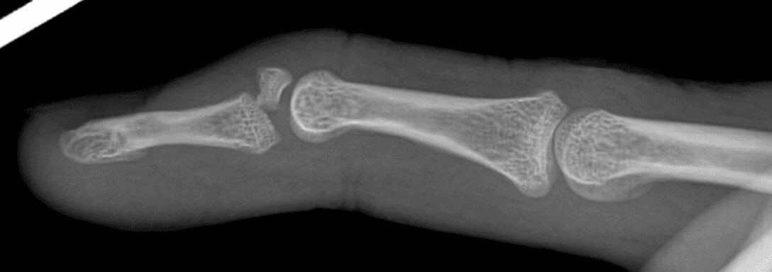
This is a bony mallet injury - avulsion of the terminal extensor tendon with a dorsal lip fragment of the distal phalanx, caused by forced flexion of an extended DIPJ.
The discriminator in this question is volar subluxation of the distal phalanx. The majority of closed bony mallet injuries - even with substantial articular involvement, heal satisfactorily with continuous DIPJ extension splinting for 6–8 weeks, and systematic reviews demonstrate no clear superiority of surgery over splinting in non-subluxated injuries. Subluxation, however, is the one widely-accepted surgical indication: without reduction the joint remains incongruent and develops post-traumatic arthritis.
The Ishiguro extension block technique is the standard approach: a K-wire is placed obliquely through the head of the middle phalanx dorsal to the avulsed fragment to act as a buttress, the DIPJ is then extended to lever the fragment into reduction, and a second trans-articular K-wire maintains the DIPJ in extension. It is closed, low-morbidity and reliably restores joint congruity.
Other answers:
Splinting alone (A) fails when subluxation is present.
Hook-plate ORIF (C) is technically demanding given the small fragment.
Trans-articular K-wiring alone (D) addresses joint position but not the displaced fragment.
Arthrodesis (E) is a salvage option.
NB - some units (in line with BSSH practice) first attempt closed reduction, and, if the joint remains subluxed, stabilise with a single trans-articular K-wire alone, reserving extension block wiring for fragments too small or comminuted to reduce indirectly. Where the fragment reduces anatomically with joint reduction, this is an accepted alternative. In this vignette, however, the persistently subluxated joint with a displaced dorsal fragment is best served by the extension block technique, which controls both the fragment and the joint, and remains the most widely examined answer.
frcs(plast) part 1 question bank
Better questions.
Better preparation.
Give yourself the months that matter. Start revising with a question bank built for the exam.